产品说明
一般描述
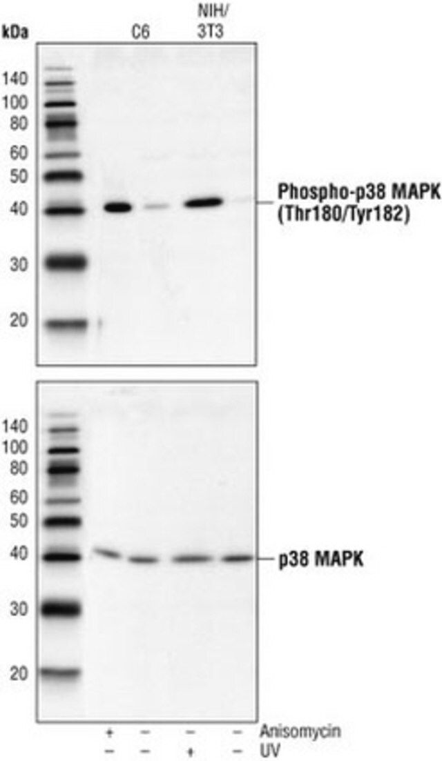
Recognizes the ~38 kDa p38 MAPK protein.
This Anti-p38 MAP Kinase (341-360) Rabbit pAb is validated for use in Flow Cytometry, Immunoblotting, Paraffin Sections for the detection of p38 MAP Kinase (341-360).
Protein A and immunoaffinity purified rabbit polyclonal antibody. Recognizes the ~38 kDa p38 MAPK protein.
免疫原
a synthetic peptide (TYDEYISFVPPPLDQEEMES) corresponding to amino acids 341-360 of human p38 MAP kinase
Human
包装
200 μL in Plastic ampoule
应用
Flow Cytometry (1:25)
Immunoblotting (1:1000)
Paraffin Sections (1:50, heat pretreatment required, see comments)
警告
Toxicity: Standard Handling (A)
外形
In 150 mM NaCl, 10 mM HEPES, 50% glycerol, 0.01% BSA, pH 7.5.
重悬
Following initial thaw, aliquot and freeze (-20°C).
其他说明
Raingeaud, J., et al. 1995. J. Biol. Chem.270, 7420.
Zervos, A.S., et al. 1995. Proc. Natl. Acad. Sci. USA92, 10531.
Han, J., et al. 1994. Science265, 808.
Lee, J.C., et al. 1994. Nature372, 739.
Rouse, J., et al. 1994. Cell78, 1027.
Pretreat paraffin sections by heating tissue in 10 mM citrate buffer, pH 6.0 for 1 min at high power followed by 9 min at medium power; keep the slides fully immersed and maintain the temperature at or just below boiling; cool the slides for 20 min at room temperature prior to staining. Variables associated with assay conditions will dictate the proper working dilution.
Recommended Protocol for Immunoblotting
Solutions and Reagents
• Transfer Buffer: 25 mM Tris base, 0.2 M glycine, 20% methanol, pH 8.5.
• SDS Sample Buffer: 62.5 mM Tris-HCl, pH 6.8, 2% SDS, 10% glycerol, 50 mM DTT, 0.1% bromophenol blue.
• 10X TBS (Tris-buffered saline): To prepare 1 liter, 24.2 g Tris base, 80 g NaCl, adjust pH to 7.6 with HCl. Dilute 1:10 for use.
• Blocking Buffer: 1X TBS, 0.1% Tween®-20 detergent with 5% non-fat dry milk.
• Primary Antibody Dilution Buffer: 1X TBS, 0.1% Tween
• Wash Buffer (TBST): 1X TBS, 0.1% Tween
Blotting Membrane
Nitrocellulose or PVDF membranes may be used.
Protein Blotting
1. Lyse cells by adding 100 ml SDS Sample Buffer and immediately scrape the cells off the plate and transfer the extract to a microfuge tube. Keep on ice.
2. Sonicate for 2 s to shear DNA and reduce sample viscosity.
3. Heat sample to 95-100°C for 5 min. Cool on ice.
4. Microcentrifuge for 5 min.
5. Load 20 ml onto SDS-PAGE gel (10 cm x 10 cm).
6. Electrotransfer to nitrocellulose membrane.
As controls, we recommend using 15 ml of phosphorylated and nonphosphorylated C-6 glioma cell extracts.
Membrane Blocking, Gel and Antibody Incubations
1. After transfer, wash membrane with 25 ml TBS for 5 min at room temperature.
2. Incubate membrane in 25 ml of Blocking Buffer for 1-3 h at room temperature or overnight at 4°C.
3. Wash 3 times for 5 min each with 15 ml TBST.
4. Incubate membrane and primary antibody (at the appropriate dilution) in 10 ml Primary Antibody Dilution Buffer with gentle agitation overnight at 4°C.
5. Wash 3 times for 5 min each with 15 ml TBST.
6. Incubate membrane with conjugated secondary antibody at the appropriate dilution in 10 ml Blocking Buffer with gentle agitation for 1 h at room temperature.
7. Wash membrane as in step 5.
Detection of Proteins
Chemiluminescence.
法律信息
CALBIOCHEM is a registered trademark of Merck KGaA, Darmstadt, Germany
TWEEN is a registered trademark of Croda International PLC
产品性质
| 生物来源 | rabbit |
| 质量水平 | 100 |
| 抗体形式 | affinity isolated antibody |
| antibody product type | primary antibodies |
| 克隆 | polyclonal |
| 形式 | liquid |
| 不包含 | preservative |
| species reactivity | mouse, rat, human |
| manufacturer/tradename | Calbiochem® |
| 储存条件 | OK to freeze avoid repeated freeze/thaw cycles |
| 同位素/亚型 | IgG |
| 运输 | wet ice |
| 储存温度 | −20℃ |
安全信息
| 储存分类代码 | 10 - Combustible liquids |
| WGK | WGK 1 |

