产品说明
一般描述
Synaptophysin (UniProt P20488; also known as Major synaptic vesicle protein p38) is encoded by the SYP gene (Gene ID 280937) in bovine species. Synaptophysin is a 38 kDa four-transmembrane glycoprotein on synaptic vesicle (SV) with both its N- and C-termini exposed cytoplasmically. Synaptophysin forms homomultimers in the SV membrane and is heavily phosphorylated on tyrosine residues in its C-terminal pentapeptide repeats. Synapthophysin is also known to interact with SV v-SNARE protein synaptobrevin II (sybII) when sybII is not engaged with other SNARE proteins, suggesting a role of synaptophysin in sybII retrieval during SV endocytosis. Consistently, synaptophysin-knockout neurons display reduced level of sybII at their nerve terminals.
特异性
Clone SY38 detects synaptophysin and stains synaptophysin-containing vesicles by targeting the flexible segment -SGGGG- in the center of the C-termimal pentapeptide repeats (Knaus, P., and Betz, H. (1990). FEBS Lett. 261(2):358-360).
免疫原
Presynaptic vesicles from bovine brain (Wiedenmann, B., and Franke, W.W. (1985). Cell. 41(3):1017-1028).
应用
Anti-Synaptophysin, clone SY38, Cat. No. MAB5258-I, is a highly specific mouse monoclonal antibody that targets Synaptophysin and has been tested in Immunocytochemistry, Immunofluorescence, Immunohistochemistry, and Western Blotting.
Research Category
Neuroscience
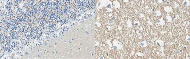
Immunohistochemistry Analysis: A 1:50 dilution from a representative lot detected Synaptophysin in human cerebellum and cerebral cortex tissue sections.
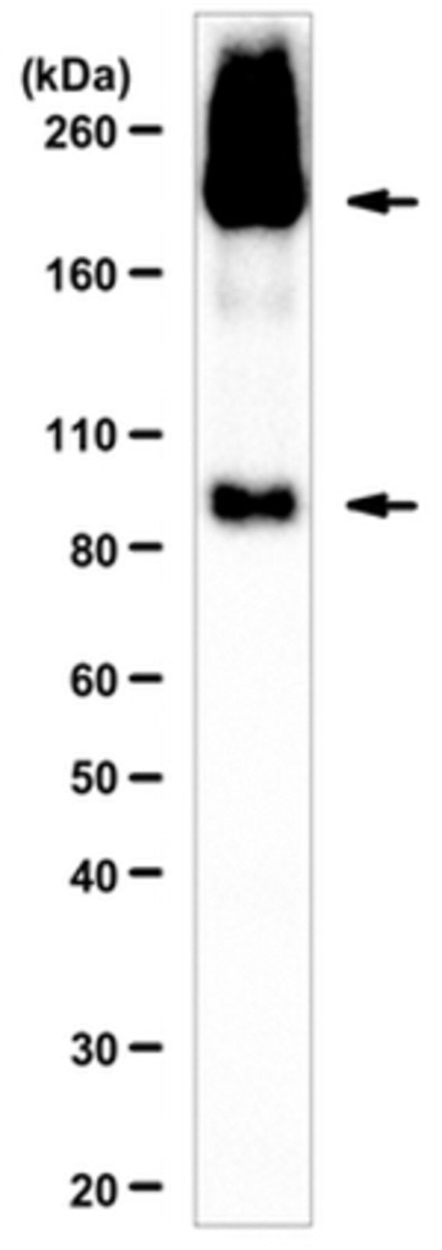
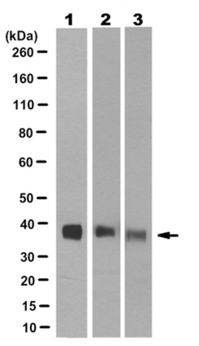
Western Blotting Analysis: A 1:1,000 dilution from a representative lot detected Synaptophysin in 10 µg of rat hippocampus and human whole brain tissue lysates.
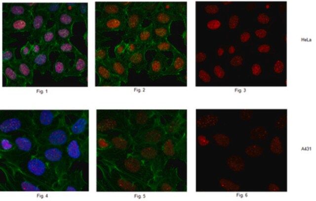
Immunocytochemistry Analysis: Representative lots immunostained presynaptic vesicles of 4% formaldehyde-fxied, 0.2% Triton X-100-permeabilzied primary mouse hippocampal neurons by fluorescent immunocytochemistry (Hu, X., et al. (2008). J. Neurosci. 28(49):13094-13105; Tarr, P.T., and Edwards, P.A. (2008). J. Lipid Res. 49(1):169-182).
Immunofluorescence Analysis: A representative lot immunostained presynaptic membrane of neurons by fluorescent immunohistochemistry staining of 4% paraformaldehyde-fixed, 0.3% Triton X-100-permeabilized, OCT-embedded rat spinal cord cryosections (Stück, E.D., et al. (2012). Neural Plast. 2012:261345).
Immunofluorescence Analysis: A representative lot immunostained synaptophysin around a large neuron in the ventral horn of the lumbar (L3/L4) segment by fluorescent immunohistochemistry staining of 2% paraformaldehyde/0.2% parabenzoquinone-fixed free-floating rat spinal cord sections (Macias, M., et al. (2009). BMC Neurosci. 10:144).
Immunofluorescence Analysis: A representative lot immunostained synaptophysin in bovine (pancreas) and human (pancreas, pheochromocytoma, and islet-cell carcinoma) frozen tissue sections (Wiedenmann, B., et al. (1986). Proc. Natl. Acad. Sci. U.S.A. 83(10):3500-3504).
Western Blotting Analysis: A representative lot detected an upregulated synaptophysin expression in human iPSCs with a 4-bp deletion in DISC1 gene (Wen, Z., et al. (2014). Nature. 515(7527):414-418).
Western Blotting Analysis: A representative lot detected synaptophysin distribution among PC12 rat pheochromocytoma cell membrane fractions (Salazar, G., et al. (2005). Mol. Biol. Cell. 16(8):3692-3704).
Western Blotting Analysis: A representative lot detected only synaptophysin recombinant contructs that contained the flexible segment -SGGGG- in the center of the c-terminal pentapeptide repeats (Knaus, P., and Betz, H. (1990). FEBS Lett. 261(2):358-360).
质量
Evaluated by Western Blotting in mouse hypothalamus tissue lysate.
Western Blotting Analysis: A 1:1,000 dilution of this antibody detected Synaptophysin in 10 µg of mouse hypothalamus tissue lysate.
目标描述
~38 kDa observed. 33.91/34.03/33.31 kDa (bovine/mouse/rat), 33.85/20.76 kDa (human isoform 1/2) calculated. Uncharacterized bands may be observed in some lysate(s).
外形
Protein G purified.
Format: Purified
Purified mouse IgG1κ in buffer containing 0.1 M Tris-Glycine (pH 7.4), 150 mM NaCl with 0.05% sodium azide.
储存及稳定性
Stable for 1 year at 2-8°C from date of receipt.
其他说明
Concentration: Please refer to lot specific datasheet.
免责声明
Unless otherwise stated in our catalog or other company documentation accompanying the product(s), our products are intended for research use only and are not to be used for any other purpose, which includes but is not limited to, unauthorized commercial uses, in vitro diagnostic uses, ex vivo or in vivo therapeutic uses or any type of consumption or application to humans or animals.
基本信息
| eCl@ss | 32160702 |
产品性质
| 质量水平 | 100 |
| 生物来源 | mouse |
| 抗体形式 | purified immunoglobulin |
| antibody product type | primary antibodies |
| 克隆 | SY38, monoclonal |
| species reactivity | mouse, rat, bovine, human |
| technique(s) | immunocytochemistry: suitable immunofluorescence: suitable immunohistochemistry: suitable (paraffin) western blot: suitable |
| 同位素/亚型 | IgG1κ |
| NCBI登记号 | NP_776388 |
| UniProt登记号 | P20488 |
| 运输 | ambient |
| Gene Information | human ... SYP(6855) |
安全信息
| 储存分类代码 | 12 - Non Combustible Liquids |
| WGK | WGK 1 |
| 闪点(F) | Not applicable |
| 闪点(C) | Not applicable |