产品说明
一般描述
Filamin is a structural protein that forms flexible cross-links between two actin filaments. Filamin is a homodimer of polypeptide chains each joined to the other at one end with an actin binding site ath the other. It is present in smooth muscle, fibroblasts, platelets and lymphocytes.
特异性
This antibody recognizes Filamin A (actin-binding protein), both the unprocessed form (270-280 kDa), and the C-terminal (90-100 kDa) calpain cleavage fragment of Filamin A (Aakhus, 1992).
免疫原
Human platelet protein corresponding to human Filamin A.
应用
This Anti-Filamin A Antibody, clone TI10, Ascites Free is validated for use in western blotting, IHC, IP, flow cytometry, immunofluorescence & ICC for the detection of Filamin A.
Research Category
Cell Structure
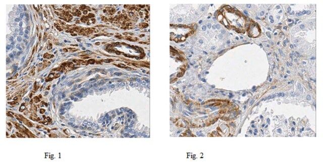
Immunohistochemistry Analysis: A 1:50 and 1:500 dilution from a representative lot detected Filamin A in human prostate and kidney tissues, respectively.
Immunohistochemistry Analysis: A representative lot from an independent laboratory detected Filamin A in human tumor tissue sections (Bedolla, R. G., et al. (2009). Clin Cancer Research. 15(3):788-796.).
Western Blotting Analysis: A representative lot from an independent laboratory detected Filamin A in human normal and tumor tissue lysates (Bedolla, R. G., et al. (2009). Clin Cancer Research. 15(3):788-796.).
Immunoprecipitation/Western Blotting Analysis: A representative lot from an independent laboratory immunoprecipitated Filamin A from transfected HEK293T cell lysates. The immunoprecipitated sample was then subjected to Western Blotting using the same lot of antibody (Nakagawa, K., et al. (2010). Biochem. J. 427(2):237–245.).
Western Blotting Analysis: A representative lot from an independent laboratory detected Filamin A in transfected U937 cell lysate (Beekman, J. M., et al. (2008). J Immunol. 180(6):3938-3945.).
Western Blotting Analysis: A representative lot from an independent laboratory detected Filamin A in prostate cancer cell lysate. (Lin, J., et al. (2007). Int. J. Cancer. 121(12):2596–2605.).
Western Blotting Analysis: A representative lot from an independent laboratory detected Filamin A in LNCap and C4-2 whole cell lysates (Wang, Y., et al. (2007). Oncogene. 26(41):6061–6070.).
Western Blotting Analysis: A representative lot from an independent laboratory detected pulled-down human Filamin A proteins. (Lad, Y., et al. (2007). EMBO J. 26(17):3993–4004.).
Western Blotting Analysis: A representative lot from an independent laboratory detected Filamin A in lysate samples from normal bladder mucosa and muscle invasive bladder tumor (Smith, S. C., et al. (2007). Clin Cancer Res. 13(13):3803-3813.).
Western Blotting Analysis: A representative lot from an independent laboratory detected Filamin A in lysate samples from human prefontal cortical tissue (Koh, P. O., et al. (2003). Ach Gen Psychiatry. 60(3):311-319.).
Flow Cytometry Analysis: A representative lot from an independent laboratory detected Filamin A in transfected U937 cells (Beekman, J. M., et al. (2008). J Immunol. 180(6):3938-3945.).
Flow Cytometry Analysis: A representative lot from an independent laboratory detected Filamin A in LAN-1 cells (Bachmann, A. S., et al. (2006) Cancer Sci. 97(12):1359–1365.).
Immunofluorescence Analysis: A representative lot from an independent laboratory detected Filamin A in clinical patient samples (Kley, R. A., et al. (2007). Brain. 130(Pt 12):3250-3264.).
Immunocytochemistry Analysis: A representative lot from an independent laboratory detected Filamin A in LAN-1 cells (Bachmann, A. S., et al. (2006) Cancer Sci. 97(12):1359–1365.).
Research Sub Category
Cytoskeleton
质量
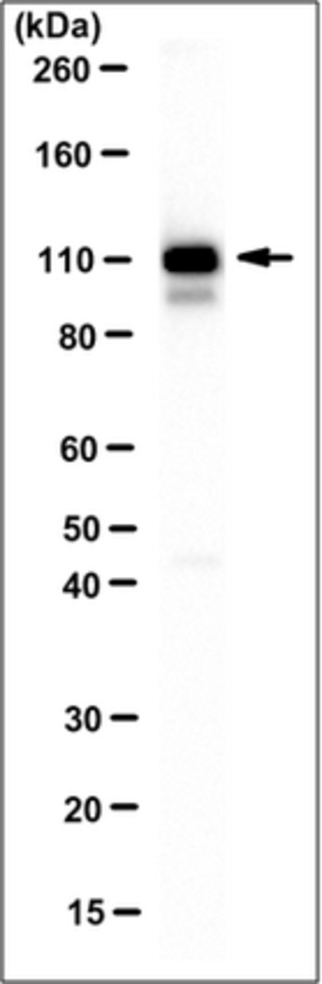
Evaluated by Western Blotting in human uterus tissue lysate.
Western Blotting Analysis: 0.5 µg/mL of this antibody detected Filamin A in 10 µg of human uterus tissue lysate.
目标描述
~110 kDa observed. Phosphorylation of this protein may cause the band to shift in Western Blotting.
外形
Purified mouse monoclonal IgG1κ in buffer containing 0.1 M Tris-Glycine (pH 7.4), 150 mM NaCl with 0.05% sodium azide.
Format: Purified
Protein G Purified
储存及稳定性
Stable for 1 year at 2-8°C from date of receipt.
免责声明
Unless otherwise stated in our catalog or other company documentation accompanying the product(s), our products are intended for research use only and are not to be used for any other purpose, which includes but is not limited to, unauthorized commercial uses, in vitro diagnostic uses, ex vivo or in vivo therapeutic uses or any type of consumption or application to humans or animals.
基本信息
| eCl@ss | 32160702 |
| NACRES | NA.41 |
产品性质
| 质量水平 | 100 |
| 生物来源 | mouse |
| 抗体形式 | purified immunoglobulin |
| antibody product type | primary antibodies |
| 克隆 | TI10, monoclonal |
| species reactivity | human |
| species reactivity (predicted by homology) | bovine (based on 100% sequence homology) |
| 浓度 | 1 mg/mL |
| technique(s) | flow cytometry: suitable immunocytochemistry: suitable immunofluorescence: suitable immunohistochemistry: suitable immunoprecipitation (IP): suitable western blot: suitable |
| 同位素/亚型 | IgG1κ |
| NCBI登记号 | NP_001104026 |
| UniProt登记号 | P21333 |
| 运输 | wet ice |
安全信息
| 储存分类代码 | 12 - Non Combustible Liquids |
| WGK | nwg |
| 闪点(F) | Not applicable |
| 闪点(C) | Not applicable |