产品说明
一般描述
Cadherin-2 (UniProt P19022; also known as CD325, CDw325, N-cadherin, Neural cadherin) is encoded by the CDH2 (also known as CDHN, NCAD) gene (Gene ID 1000) in human. Cadherins constitute a family of calcium-dependent cell-cell adhesion proteins that play important roles in the embryonic development and maintenance of normal tissue architecture. Cadherins are composed of an extracellular domain (a.a. 160-724 of human N-cadherin) with five homologous repeats that mediates adhesion, a single pass transmembrane domain (a.a. 725-745 of human N-cadherin), and a conserved cytoplasmic domain (a.a. 746-906 of human N-cadherin) that interacts with catenins to link cadherins to the actin cytoskeleton. In addition, a known Src substrate p120ctn also modulate the strength of cadherin-dependent adhesion by interacting with cadherins at their intracellular juxtamembrane domain. Cadherins are synthesized as precursor proteins that must be proteolytically cleaved to generate functional, mature proteins. Newly synthesized proN-cadherin (a.a. 1-906) is phosphorylated and proteolytically processed prior to transport to the plasma membrane. In addition, Plakoglobin (gamma-catenin) and beta-catenin associate only with phosphorylated proN-cadherin, whereas p120ctn can associate with both phosphorylated and non-phosphorylated proN-cadherin. The N-terminal signal and propeptide (a.a. 1-25 and 26-159 of of human N-cadherin) region is proteolytically removed and a core N-cadherin-catenin complex is assembled in the endoplasmic reticulum or Golgi compartment prior to localization at the plasma membrane where linkage to the actin cytoskeleton can be established.
特异性
Clone 13A9 recognizes N-cadherin, but not P-, E-, or M-cadherin (Knudsen, K.A., et al. (1995). J. Cell Biol. 130(1):67-77).
免疫原
Epitope: Cytoplasmic domain.
Bacterially expressed human N-cadherin cytoplasmic domain MBP fusion protein (Knudsen, K.A., et al. (1995). J. Cell Biol. 130(1):67-77).
应用
Research Category
Cell Structure
Immunohistochemistry Analysis: A representative lot immunostained the extracellular matrix of the stable plaques and in the fibrous cap region rich in vascular smooth muscle cells (VSMCs) using patients-derived paraffin-embedded internal carotid artery tissue sections (Musumeci, G., et al. (2014). Histol. Histopathol. 29(6):707-719).
Immunohistochemistry Analysis: A representative lot detected strong N-cadherin immunoreactivity in paraffin-embedded rectal cancer (RC) tissues with positive regional lymph node metastasis (RLNM) status, while only weak N-cadherin immunoreactivity was detected in RC with negative RLNM, and no N-cadherin staining was seen in normal colorectal epithelium (Fan, X.J., et al. (2012). Br. J. Cancer. 106(11):1735-1741).
Immunohistochemistry Analysis: A representative lot detected N-cadherin immunoreactivity in formalin-fixed, paraffin-embedded hepatocellular carcinoma (HCC) tissue sections. A significant inverse correlation was found between RUNX3 and N-cadherin expression levels (Tanaka, S., et al. (2012). Int. J. Cancer. 131(11):2537-2546).
Western Blotting Analysis: A representative lot detected an upregulated N-cadherin expression in CCL185 carcinoma cells following transient Epstein-Barr virus (EBV) infection. The EMT-like phenotype remained even after viral loss by culture selection pressure withdrawal (Queen, K.J., et al. (2013). Int. J. Cancer. 132(9):2076-2086).
Western Blotting Analysis: A representative lot detected N-cadherin in Hep3B, Huh7, HLF and SK-Hep1 human hepatocellular carcinoma (HCC) cell lysates (Tanaka, S., et al. (2012). Int. J. Cancer. 131(11):2537-2546).
Western Blotting Analysis: A representative lot detected both the unprocessed (pro-) and processed (mature) forms of N-cadherin in HeLa cell lysate (Wahl, J.K. 3rd., et al. (2003). J. Biol. Chem. 278(19):17269-17276).
Western Blotting Analysis: A representative lot detected N-cadherin in WI-38 human fibroblast lysate, but not in JAr human placental choriocarcinoma cell lysate (Knudsen, K.A., et al. (1995). J. Cell Biol. 130(1):67-77).
Immunocytochemistry Analysis: A representative lot detected N-cadherin immunoreactivity localized primarily at the cell-cell borders by fluorescent immunocytochemistry staining of 1% paraformaldehyde-fixed, methanol-permeabilized HeLa cells (Wahl, J.K. 3rd., et al. (2003). J. Biol. Chem. 278(19):17269-17276).
Immunocytochemistry Analysis: A representative lot detected N-cadherin immunoreactivity colocalized with those of alpha- and beta-catenin by dual fluorescent immunocytochemistry staining of fixed WI-38 human fibroblasts (Knudsen, K.A., et al. (1995). J. Cell Biol. 130(1):67-77).
Immunoprecipitation Analysis: Representative lots co-immunoprecipitated alpha-catenin, beta-catenin, and plakoglobin with N-cadherin from WI-38 human fibroblast and HeLa cell lysates (Wahl, J.K. 3rd., et al. (2003). J. Biol. Chem. 278(19):17269-17276; Knudsen, K.A., et al. (1995). J. Cell Biol. 130(1):67-77).
Detect N-cadherin using this Anti-N-Cadherin Antibody, clone 13A9 validated for use in Immunocytochemistry, Immunohistochemistry, Immunoprecipitation, and Western Blotting.
Research Sub Category
Adhesion (CAMs)
质量
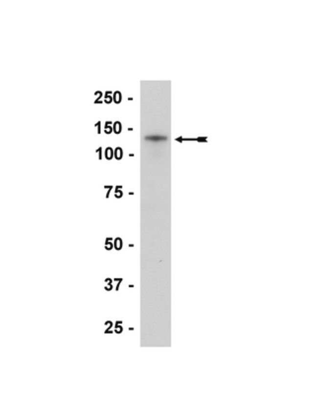
Evaluated by Western Blotting in HeLa cell lysate.
Western Blotting Analysis: A 1:1000-5000 dilution of this hybridoma culture supernatant detected N-cadherin in HeLa cell lysate.
目标描述
~140 kDa observed. Target band size appears larger than the calculated molecular weights of 82.03 kDa (mature) and 99.81/97.04 kDa (isoform 1/2 pro-form) due to posttranslational glycosylation and phosphorylation.
联系
Replaces: 04-1126
外形
Mouse monoclonal immunoglobulin hybridoma culture supernatant containing 0.05% sodium azide before the addition of glycerol to 30%.
Unpurified
储存及稳定性
Maintain for 2 years at -20°C from date of shipment. Aliquot to avoid repeated freezing and thawing. For maximum recovery of product, centrifuge the original vial after thawing and prior to removing the cap.
分析说明
Control
HeLa cell lysate
法律信息
UPSTATE is a registered trademark of Merck KGaA, Darmstadt, Germany
免责声明
Unless otherwise stated in our catalog or other company documentation accompanying the product(s), our products are intended for research use only and are not to be used for any other purpose, which includes but is not limited to, unauthorized commercial uses, in vitro diagnostic uses, ex vivo or in vivo therapeutic uses or any type of consumption or application to humans or animals.
基本信息
| eCl@ss | 32160702 |
产品性质
| 质量水平 | 100 |
| 生物来源 | mouse |
| 抗体形式 | culture supernatant |
| antibody product type | primary antibodies |
| 克隆 | 13A9, monoclonal |
| species reactivity | human |
| 包装 | antibody small pack of 25 μL |
| manufacturer/tradename | Upstate® |
| technique(s) | immunocytochemistry: suitable immunohistochemistry: suitable immunoprecipitation (IP): suitable western blot: suitable |
| NCBI登记号 | NM_001792 |
| UniProt登记号 | P19022 |
| 运输 | ambient |
| Gene Information | human ... CDH2(1000) |