产品说明
一般描述
Claudin-1 (UniProt O95832; also known as Senescence-associated epithelial membrane protein) is encoded by the CLDN1 (also known as CLD1, ILVASC, NISCH, SEMP1) gene (ORF UNQ481/PRO944; Gene ID 9076) in human. Tight junctions (TJs) are highly specialized membrane barriers that block the free passage of small molecules, microorganisms, and cells across the paracellular space, as well as provide a physical barrier between the apical and basolateral membrane domains of a polarized epithelial cell. Claudins are TJ proteins whose main function is to regulate the passage of ions across the paracellular space. There exist at least 27 human claudins with differential epithelial expression patterns, allowing tissue-specific paracellular transport of ions. Claudin-1 is expressed on apical and basolateral surfaces of hepatocytes in normal liver tissue. Claudin-1 is a known host factor for hepatitis C virus (HCV) entry via interaction with CD81 and cell-to-cell HCV transmission. Claudin-1 is a four-transmembrane (a.a. 8-28, 82-102, 116-136, 164-184) protein, having both its N- and C-terminal ends exposed cytoplasmically (a.a. 1-7 & 185-211).
特异性
Clone 7A5 specifically recognized human claudin-1/CLDN1 by targeting an epitope in the second extracellular loop and prevented claudin-1-dependent hepatitis C virus (HCV) entry into host cells. Clone 7A5 does not react with human claudin-2, -4, -5, -6, -7, or -9, nor mouse claudin-1 (Fukasawa, M., et al. (2015). J. Virol. 89(9):4866-4879).
免疫原
Recombinant human Claudin-1/CLDN1.
Epitope: Second extracellular loop.
应用
Research Category
Cell Structure
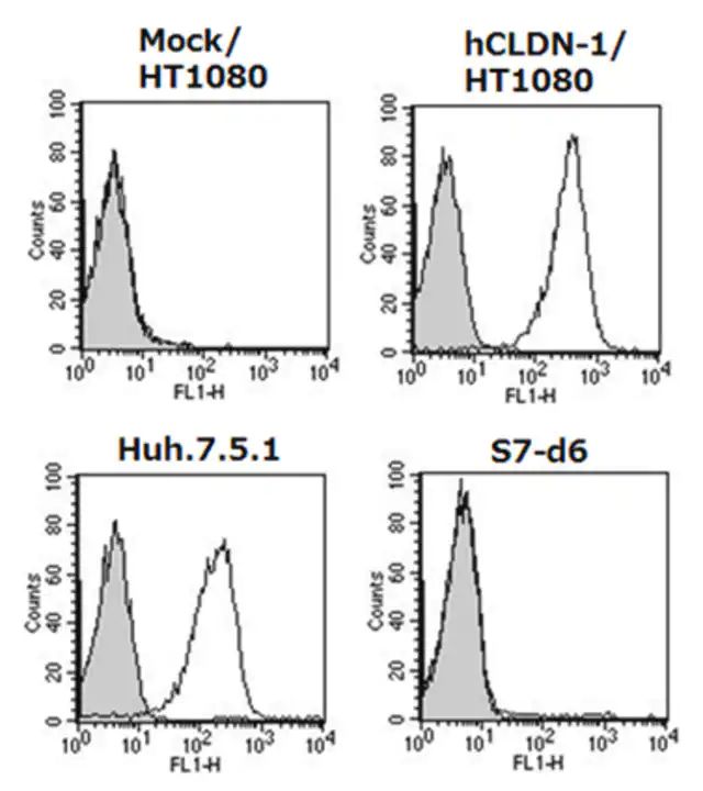
Flow Cytometry Analysis: 5.0 µg/mL from a representative lot immunostained HT1080 cells expressing exogenously transfected human claudin-1/CLDN1 and Huh-7.5.1 human hepatoma cells, but not mock-transfected HT1080 cells or non-claudin-1-expressing S7-d6 cells (Courtesy of Professor Masuo Kondoh, PhD, Osaka University, Japan).
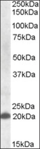
Western Blotting Analysis: 0.5 µg/mL from a representative lot detected exogenously expressed claudin-1 in CLDN1-transfected, but not mock-transfected, HT1080 cells (Courtesy of Professor Masuo Kondoh, PhD, Osaka University, Japan).
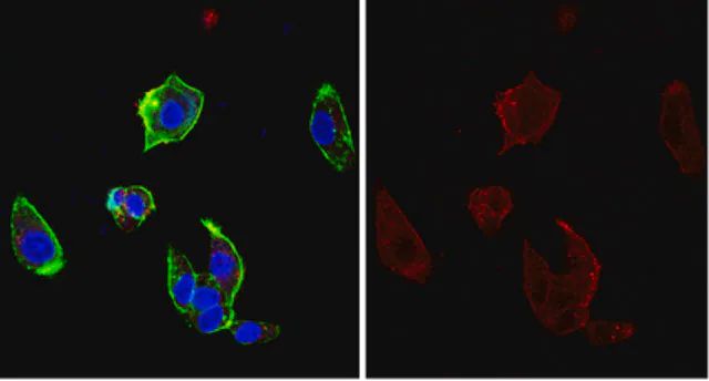
Immunocytochemistry Analysis: A representative lot immunostained the surface of Huh-7.5.1 human hepatoma cells, but not the non-claudin-1-/CLDN1-expressing S7-A cells (Fukasawa, M., et al. (2015). J. Virol. 89(9):4866-4879).
Flow Cytometry Analysis: A representative lot specifically immunostained HT1080 cells expressing exogenously transfected human claudin-1 (CLDN1), but not HT1080 cells expressing human claudin-2, -4, -5, -6, -7, or -9, nor L cells expressing mouse claudin-1 (Fukasawa, M., et al. (2015). J. Virol. 89(9):4866-4879).
Flow Cytometry Analysis: A representative lot immunostained HEK293T transfectants expressing FLAG-tagged human claudin-1/CLDN1 and human-mouse claudin-1 chimeras with the second human extracellular loop (EL2), but not chimeras with the mouse EL2. M152L, but not V155I, mutation in human EL2 abolished the immunoreactivity (Fukasawa, M., et al. (2015). J. Virol. 89(9):4866-4879).
Western Blotting Analysis: A representative lot detected FLAG-tagged human claudin-1/CLDN1 and human-mouse claudin-1 chimeras with the second human extracellular loop (EL2), but not chimeras with the mouse EL2. M152L, but not V155I, mutation in the second human extracellular loop abolished the immunoreactivity (Fukasawa, M., et al. (2015). J. Virol. 89(9):4866-4879).
ELISA Analysis: A representative lot detected claudin-1/CLDN1 immunoreactivity in 3.7% formaldehyde-fixed Huh-7.5.1 human hepatoma cells by "cell ELISA" (Fukasawa, M., et al. (2015). J. Virol. 89(9):4866-4879).
Neutralization Analysis: A representative lot inhibited HCV infection of cultured Huh-7.5.1 human hepatoma cells in vitro and of human liver-chimeric mice in vivo (Fukasawa, M., et al. (2015). J. Virol. 89(9):4866-4879).
Research Sub Category
Infectious Diseases - Viral
This Anti-Claudin-1 Antibody, clone 7A5 is validated for use in Immunocytochemistry, Western Blotting, Flow Cytometry for the detection of CLDN1.
质量
Evaluated by Immunocytochemistry in HepG2 cells.
Immunocytochemistry Analysis: 10 µg/mL of this antibody detected Claudin-1/CLDN1 in HepG2 cells.
目标描述
~20 kDa observed. 22.74 kDa calculated.
外形
Protein G purified.
Format: Purified
Purified mouse monoclonal IgG1κ antibody in PBS without preservatives.
储存及稳定性
Stable for 1 year at -20°C from date of receipt.
Handling Recommendations: Upon receipt and prior to removing the cap, centrifuge the vial and gently mix the solution. Aliquot into microcentrifuge tubes and store at -20°C. Avoid repeated freeze/thaw cycles, which may damage IgG and affect product performance.
其他说明
Concentration: Please refer to lot specific datasheet.
免责声明
Unless otherwise stated in our catalog or other company documentation accompanying the product(s), our products are intended for research use only and are not to be used for any other purpose, which includes but is not limited to, unauthorized commercial uses, in vitro diagnostic uses, ex vivo or in vivo therapeutic uses or any type of consumption or application to humans or animals.
基本信息
| eCl@ss | 32160702 |
| NACRES | NA.41 |
产品性质
| 质量水平 | 100 |
| 生物来源 | mouse |
| 抗体形式 | purified immunoglobulin |
| antibody product type | primary antibodies |
| 克隆 | 7A5, monoclonal |
| species reactivity | human |
| should not react with | mouse |
| technique(s) | flow cytometry: suitable immunocytochemistry: suitable western blot: suitable |
| 同位素/亚型 | IgG1κ |
| NCBI登记号 | NP_066924 |
| UniProt登记号 | O95832 |
| 运输 | dry ice |
| Gene Information | human ... CLDN1(9076), NISCH(11188) |
安全信息
| 储存分类代码 | 12 - Non Combustible Liquids |
| WGK | WGK 2 |
| 闪点(F) | Not applicable |
| 闪点(C) | Not applicable |