产品说明
一般描述
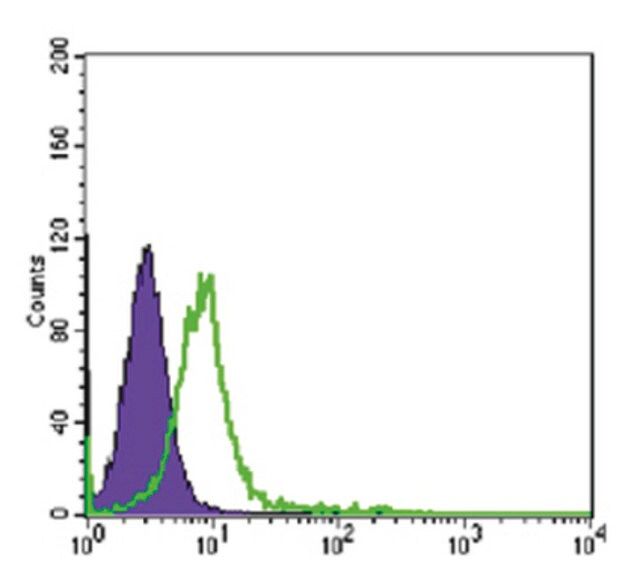
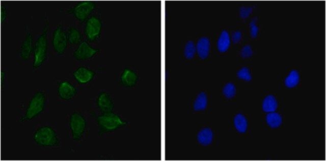
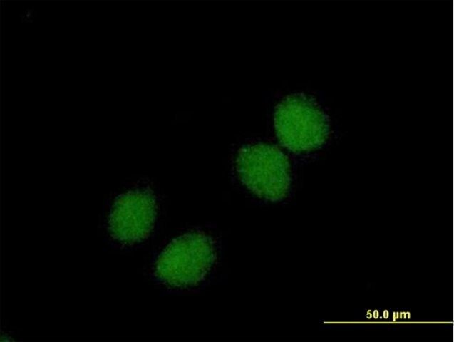
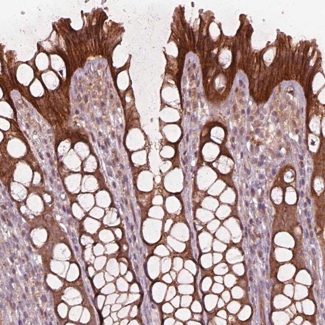
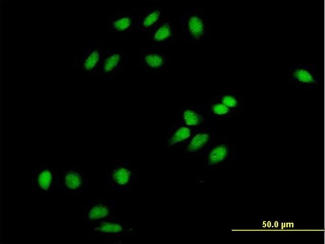
Chromodomain-helicase-DNA-binding protein 3 (CHD-3) or alternatively known as ATP-dependent helicase CHD3, Mi-2 autoantigen 240kDa protein, Mi2-alpha, Zinc finger helicase (hZFH) and encoded by the human gene CHD3 is a member of the CHD family of proteins which are characterized by the presence of a chromo (chromatin organization modifier) domains and SNF2-related helicase/ATPase domains. CHD3 is one member of a large histone deacetylase complex called NuRD. CHD3 is one of the ATPases that form part of the NuRD complex. The NuRD complex operates in the remodeling of chromatin by deacetylating histones and thus typically functions as a transcriptional repressor, though NuRD can also act as an activator depending upon the system studied. CHD3 via the NuRD complex also interacts with HDAC1 and HDAC2. CHD3 is also required for anchoring of the centrosomal protein pericentrin and for the organization of the spindle and centrosome during cell division. It is typically located in the nucleus but CHD3 is seen cytoplasmically during mitosis associated with the centrosomes. CHD3 is expressed by most cells and tissues. Interestingly CHD3 is also a hallmark auto antigen of MI-2 dermatomyositis. EMD-Millipore’s Anti-CHD3 mouse monoclonal antibody has been tested in western blot against HeLa, K562, Jurkat, NTERA-2, HEK293, Raji cells lysates and mouse brain tissue lysate and in paraffin embedded immunohistochemistry on human colon cancer tissue sections. The antibody has also been successfully tested in fluorescent immunocytochemistry on HeLa cells in culture and by flow cytometry on K562 cells in culture.
免疫原
Purified recombinant fragment of human CHD3 expressed in E. Coli.
应用
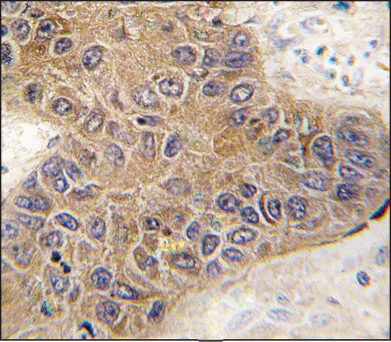
Immunohistochemistry Analysis: A 1:200-1,000 dilution from a representative lot detected CHD3 in colon cancer tissue.
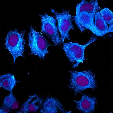
Immunofluorescence Analysis: A 1:200-1,000 dilution from a representative lot detected CHD3 in HeLa cells.
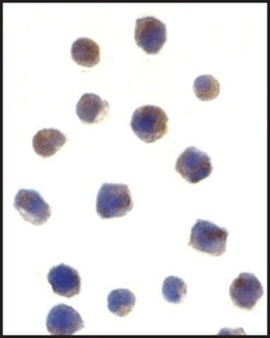
Flow cytometry analysis: A 1:200-400 dilution from a representative lot detected CHD3 in K562 cells.
Optimal working dilutions must be determined by end user.
Research Category
Neuroscience
Research Sub Category
Developmental Neuroscience
Detect CHD-3 using this mouse monoclonal antibody, Anti-CHD3 Antibody, clone 2G4 validated for use in western blotting, IHC, Immunofluorescence & Flow Cytometry.
质量
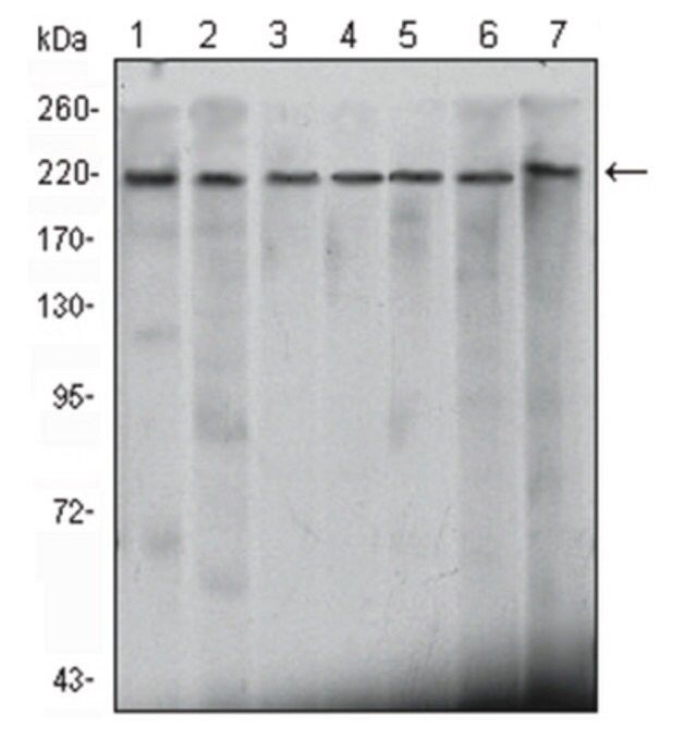
Evaluated by Western Blotting in mouse brain, HeLa, K562, Jurkat, NTERA-2, HEK293, Raji lysates.
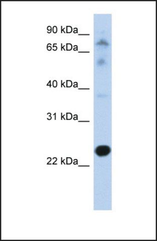
Western Blotting Analysis: A 1:500-2,000 dilution of this antibody detected CHD3 in mouse brain, HeLa, K562, Jurkat, NTERA-2, HEK293, Raji lysates.
目标描述
~220 kDa observed. Uncharacterized bands may appear in some lysate(s).
联系
Replaces: MABE482
外形
Unpurified
Mouse monoclonal IgG1 ascitic fluid containing up to 0.1% sodium azide.
储存及稳定性
Stable for 1 year at -20°C from date of receipt.
Handling Recommendations: Upon receipt and prior to removing the cap, centrifuge the vial and gently mix the solution. Aliquot into microcentrifuge tubes and store at -20°C. Avoid repeated freeze/thaw cycles, which may damage IgG and affect product performance.
分析说明
Control
mouse brain, HeLa, K562, Jurkat, NTERA-2, HEK293, Raji lysates
免责声明
Unless otherwise stated in our catalog or other company documentation accompanying the product(s), our products are intended for research use only and are not to be used for any other purpose, which includes but is not limited to, unauthorized commercial uses, in vitro diagnostic uses, ex vivo or in vivo therapeutic uses or any type of consumption or application to humans or animals.
基本信息
| eCl@ss | 32160702 |
| NACRES | NA.41 |
产品性质
| 质量水平 | 100 |
| 生物来源 | mouse |
| 抗体形式 | ascites fluid |
| antibody product type | primary antibodies |
| 克隆 | 2G4, monoclonal |
| species reactivity | mouse, human |
| technique(s) | flow cytometry: suitable immunofluorescence: suitable immunohistochemistry: suitable western blot: suitable |
| 同位素/亚型 | IgG1 |
| UniProt登记号 | Q12873 |
| 运输 | wet ice |
安全信息
| 储存分类代码 | 12 - Non Combustible Liquids |
| WGK | nwg |
| 闪点(F) | Not applicable |
| 闪点(C) | Not applicable |

