
产品说明
一般描述
Actin, cytoplasmic 1 (UniProt P60709; also known as Beta-actin) is encoded by the ACTB gene (Gene ID 60) in human. Actins are globular multi-functional proteins that serve as the basic building blocks of cytoskeletal microfilaments and are among the most conserved eukaryotic proteins. Six actin types exist, skeletal muscle alpha-actin is encoded by the ACTA1 gene, smooth muscle alpha-actin by the ACTA2 gene, cytoplasmic beta-actin by the ACTB gene, cardiac muscle alpha-actin by the ACTC1 gene, cytoplasmic gamma-actin by the ACTG1 gene, and smooth muscle gamma-actin by the ACTG2 (a.k.a. ACTA3) gene. Although actins show >90% overall sequence homology, isoforms do show spatial, temporal, and tissue-specific expression patterns and only 50-60% homology is found in their 18 N-terminal residues. Cytoplasmic β and γ-actins, are thought to be present in all cells, while the other four actin isoforms are typically found in specific adult muscle tissue types. Actins exist in a variety of structural states, depending on the specific ionic conditions or the interaction with ligand proteins. The oligomeric and polymeric forms that actin molecules assume are dependent on the distinct conformations they adopt.
特异性
Clone 4C2 detected BSA conjugated with beta-actin N-terminal peptide, but not BSA conjugated with N-terminal peptides derived from the 5 other actin types (Dugina, V., et al. (2009). J. Cell Sci. 122(Pt 16):2980-2988).
免疫原
KLH-conjugated linear peptide corresponding to the the N-terminal sequence of human beta-Actin.
应用
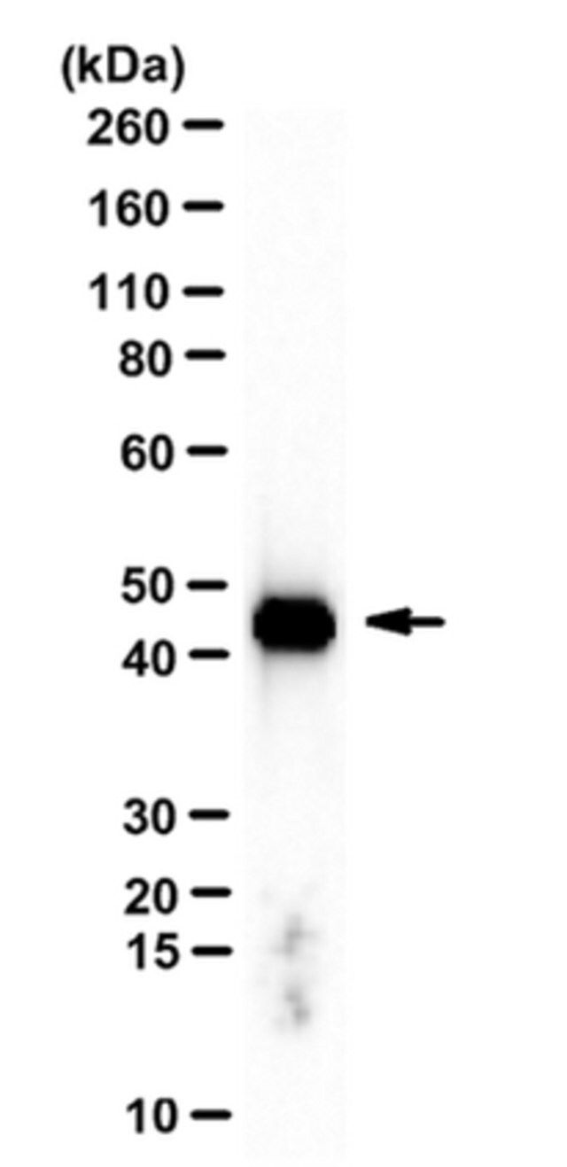
Western Blotting Analysis: 0.5 µg/mL from a representative lot detected beta-Actin in 10 µg of NIH/3T3 cell lysate.
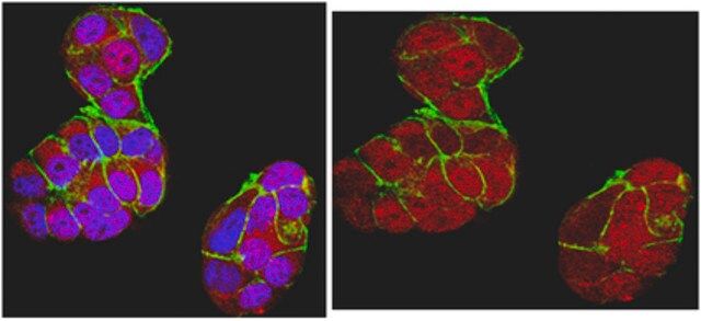
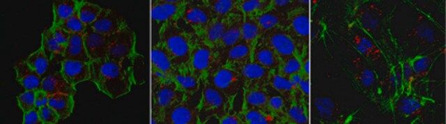
Immunocytochemistry Analysis: 5.0 µg/mL from a representative lot detected beta-Actin in HUVECs, A431 and HeLa cells.
Western Blotting Analysis: A representative lot detected downregulated beta-actin levels in stimulated (by A23187, TRAP-6, TNF, LPS, or IFN-γ) human cerebral microvascular endothelial D3 cells (hCMEC/D3) and their microparticles (MPs) when compared with unstimulated hCMEC/D3 and their MPs (Latham, S.L., et al. (2013). FASEB J. 27(2):672-683).
Western Blotting Analysis: A representative lot detected siRNA-mediated downregulation of beta-actin in A549 human lung carcinoma cells (Miazza, V., et al. (2011). Virology. 410(1):7-16).
Western Blotting Analysis: A representative lot detected beta-actin, but not cytoplasmic gamma-actin separated by 2-D gel electrophoresis of purified chicken gizzard actins or total protein extracts from human subcutaneous fibroblasts (HSCFs), canine MDCK cells, and rat aorta tissue (Dugina, V., et al. (2009). J. Cell Sci. 122(Pt 16):2980-2988).
Western Blotting Analysis: A representative lot detected BSA conjugated with beta-actin N-terminal peptide, but not BSA conjugated with N-terminal peptides derived from the other 5 actin types (Dugina, V., et al. (2009). J. Cell Sci. 122(Pt 16):2980-2988).
Immunocytochemistry Analysis: A representative lot detected TNF-stimulated localization of β-actin into thick, intensely staining stress fibers prominent at the basal surface of of human cerebral microvascular endothelial D3 cells (hCMEC/D3). Rho kinase inhibitor Y-27632 (Cat. No. 688000) treatment suppressed TNF-induced β-actin stress fiber formation (Latham, S.L., et al. (2013). FASEB J. 27(2):672-683).
Immunocytochemistry Analysis: A representative lot detected a drastic subcellular redistribution of beta-actin following Sendai virus infection of polarized Madin-Darby canine kidney (MDCK) epithelial cells by fluorescent immunocytochemistry staining of paraformaldehyde-fixed, methanol-treated cells (Miazza, V., et al. (2011). Virology. 410(1):7-16).
Immunocytochemistry Analysis: A representative lot detected beta-actin subcellular localization distinct from that of cytoplasmic gamma-actin in both spreading and stationary cells by fluorescent immunocytochemistry, using paraformaldehyde-fixed, methanol-treated HSCF human subcutaneous fibroblasts, HaCaT human keratinocytes, WI38 human embryonic fibroblasts,and Madin-Darby canine kidney (MDCK) cells (Dugina, V., et al. (2009). J. Cell Sci. 122(Pt 16):2980-2988).
This Anti-beta-Actin Antibody, clone 4C2 is validated for use in Western Blotting, Immunocytochemistry for the detection of beta-Actin.
质量
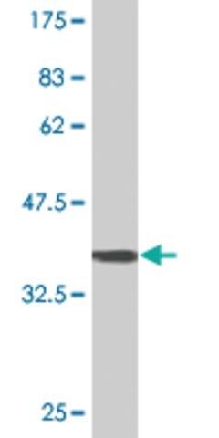
Evaluated by Western Blotting in HeLa cell lysate.
Western Blotting Analysis: 0.5 µg/mL of this antibody detected beta-Actin in 10 µg of HeLa cell lysate.
目标描述
~39-45 kDa observed. Uncharacterized band(s) may appear in some lysates.
外形
Protein G Purified
Format: Purified
其他说明
Concentration: Please refer to lot specific datasheet.
免责声明
Unless otherwise stated in our catalog or other company documentation accompanying the product(s), our products are intended for research use only and are not to be used for any other purpose, which includes but is not limited to, unauthorized commercial uses, in vitro diagnostic uses, ex vivo or in vivo therapeutic uses or any type of consumption or application to humans or animals.
基本信息
| eCl@ss | 32160702 |
| NACRES | NA.41 |
产品性质
| 质量水平 | 100 |
| 生物来源 | mouse |
| 抗体形式 | purified immunoglobulin |
| antibody product type | primary antibodies |
| 克隆 | 4C2, monoclonal |
| species reactivity | chicken, rat, mouse, human |
| species reactivity (predicted by homology) | mammals (based on 100% sequence homology) |
| technique(s) | immunocytochemistry: suitable western blot: suitable |
| 同位素/亚型 | IgG1κ |
| NCBI登记号 | NP_001092.1 |
| UniProt登记号 | P60709 |
| 运输 | wet ice |
安全信息
| 储存分类代码 | 12 - Non Combustible Liquids |
| WGK | WGK 1 |
| 闪点(F) | Not applicable |
| 闪点(C) | Not applicable |