产品说明
一般描述
Wilms tumor protein (UniProt P19544; also known as WT33) is encoded by the WT1 (also known as DDS, FS, MEACHS, MESOM, NPHS4, WAGR) gene (Gene ID 7490) in human. The Wilms’ tumor gene WT1 was originally identified in the childhood kidney cancer Wilms’ tumor. The N-terminal region of WT1 protein contains a proline-rich region (a.a. 27-83) involved in transcriptional regulation, self-association, and RNA recognition, while its C-terminal region contains four zinc fingers (a.a 323-347, 353-377, 383-405, 414-438) that mediate DNA and RNA binding. The zinc finger domain of WT1 can bind to GC-rich sequences, such as the EGR-1 consensus sequence (5’-GCG(T/G)GGGCG-3’), the WTE motif (5′-GCGTGGGAGT-3′), or (TCC)n motif. Many genes responsible for cell growth and apoptosis, such as Bcl-2, Bcl-xL, BFL1, and c-myc, have been identified as downstream targets of WT1. There are four major alternatively spliced WT1 isoforms resulting from splicing at either or both of exon 5 (17AA) and exon 9 (KTS). All four major WT1 isoforms are overexpressed in leukemia and solid tumors and play oncogenic roles such as inhibition of apoptosis, and promotion of cell proliferation, migration and invasion.
应用
This Anti-Wilms′ Tumor Antibody, NT clone 6F-H2, Ascites Free is validated for use in Immunocytochemistry, Immunoprecipitation, Immunofluorescence, Immunohistochemistry (Paraffin), and Western Blotting for the detection of Wilms′ tumor protein.
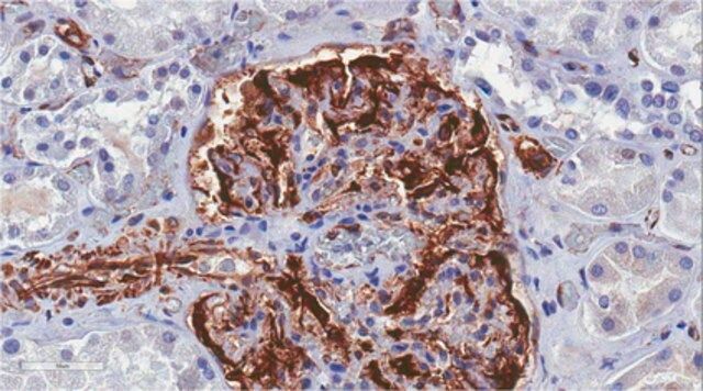
Immunohistochemistry Analysis: An 1:250 dilution from a representative lot detected Wilms tumor protein in human kidney tissue.
Immunoprecipitation Analysis: A representative lot co-immunoprecipitated CRE-binding protein/CBP together with Wilms tumor protein WT1 from the lysate of a T-SV40 immortalized human glomerular epithelial cell (HGEC) line (Drossopoulou, G.I., et al. (2009). Am. J. Physiol. Renal Physiol. 297(3):F594-F603).
Western Blotting Analysis: A representative lot detected Wilms tumor protein WT1 in the CRE-binding protein/CBP immunoprecipitate obtained from the lysate of a T-SV40 immortalized human glomerular epithelial cell (HGEC) line (Drossopoulou, G.I., et al. (2009). Am. J. Physiol. Renal Physiol. 297(3):F594-F603).
Western Blotting Analysis: A representative lot detected Wilms tumor protein WT1 in lysates from mouse E15.5 embryonic kidney and human melanoma cell lines A375, SK-MEL-28, and WM-266-4 (Wagner, N., et al. (2008). Pflugers Arch. Eur. J. Physiol. 455(5):839-847).
Immunocytochemistry Analysis: A representative lot immunostained the nucleus of methanol-fixed human melanoma A375 cells by fluorescent immunocytochemistry (Wagner, N., et al. (2008). Pflugers Arch. Eur. J. Physiol. 455(5):839-847).
Immunofluorescence Analysis: A representative lot immunostained the PCNA-positive nuclei of proliferating cells in formalin-fixed, paraffin-embedded human melanoma tissue sections by fluorescent immunohistochemistry (Wagner, N., et al. (2008). Pflugers Arch. Eur. J. Physiol. 455(5):839-847).
Immunohistochemistry Analysis: A representative lot immunostained glomeruli in formalin-fixed, paraffin-embedded normal human kidney and Wilms′ tumor sections (Wagner, N., et al. (2008). Pediatr. Nephrol. 23(9):1445-1453).
Immunohistochemistry Analysis: A representative lot detected vascular WT1 expression in 95% of 113 paraffin-embedded tumour tissues of various types. In most cases, nuclear WT1 staining of endothelial cells was seen (Wagner, N., et al. (2008). Oncogene. 27(26):3662-3672).
Immunohistochemistry Analysis: A representative lot immunostained the nucleus of perifollicular fibroblasts at the hair follicle in formalin-fixed, paraffin-embedded normal human skin sections. Most common melanocytic nevi do not express WT1, whereas Spitz nevi and dysplastic nevi show cytoplasmic WT1 staining. (Wagner, N., et al. (2008). Pflugers Arch. Eur. J. Physiol. 455(5):839-847).
质量
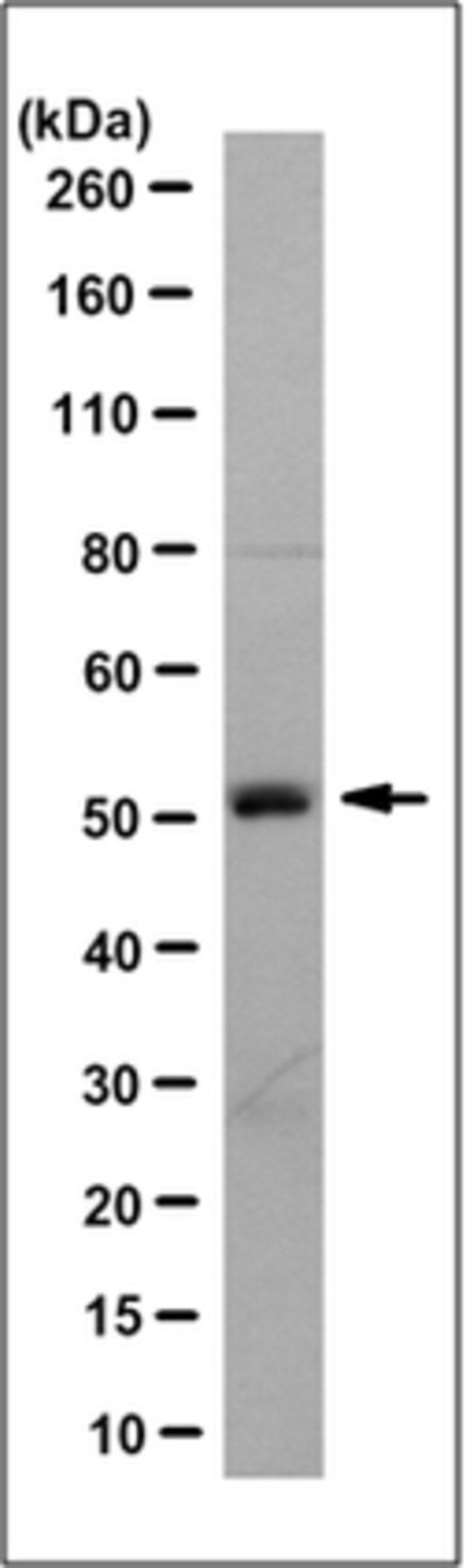
Evaluated by Western Blotting in Jurkat cell lysate.
Western Blotting Analysis: 1.0 µg/mL of this antibody detected Wilms tumor protein in 10 µg of Jurkat cell lysate.
目标描述
~52 kDa observed. 49.19 kDa (isoform 1), 47.20 kDa (isoform 2), 47.51 kDa (isoform 3), 48.87 kDa (isoform 4), 34.45 kDa (isoform 5), 56.88 kDa (isoform 6), 55.21 kDa (isoform 7), 33.09 kDa (isoform 8) calculated. Uncharacterized band(s) may appear in some lysates.
外形
Protein G purified.
Format: Purified
其他说明
Concentration: Please refer to lot specific datasheet.
免责声明
Unless otherwise stated in our catalog or other company documentation accompanying the product(s), our products are intended for research use only and are not to be used for any other purpose, which includes but is not limited to, unauthorized commercial uses, in vitro diagnostic uses, ex vivo or in vivo therapeutic uses or any type of consumption or application to humans or animals.
基本信息
| eCl@ss | 32160702 |
| NACRES | NA.41 |
产品性质
| 质量水平 | 100 |
| 生物来源 | mouse |
| 抗体形式 | purified immunoglobulin |
| antibody product type | primary antibodies |
| 克隆 | 6F-H2, monoclonal |
| species reactivity | mouse, human |
| technique(s) | immunocytochemistry: suitable immunofluorescence: suitable immunohistochemistry: suitable (paraffin) immunoprecipitation (IP): suitable western blot: suitable |
| 同位素/亚型 | IgG1κ |
| NCBI登记号 | NP_000369 |
| UniProt登记号 | P19544 |
| 运输 | wet ice |
| Gene Information | human ... WT1(7490) |
安全信息
| 储存分类代码 | 12 - Non Combustible Liquids |
| WGK | WGK 1 |
| 闪点(F) | Not applicable |
| 闪点(C) | Not applicable |