产品说明
一般描述
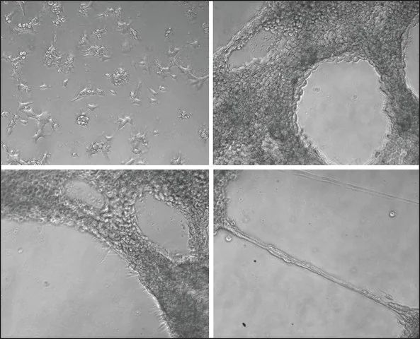
HMT-3522 T4-2, also known as HMT-3522/mt-1, is a subline that has been derived from HMT-3522. The parent line was established from a benign breast tumor of a 48 year old woman and has undergone spontaneous malignant transformation. It was grown on collagen in serum-free media without antibiotics from explantation onwards. A mutation at codon 179 of the p53 gene was detected during establishment of the parent line at passage 25-30. The subline S1 is a continuation of this line from passage 34 (ECACC catalogue no. 98102210) growing without collagen. Subline S2 (ECACC catalogue no. 98102211) has been isolated by culturing S1 cells from passage 118 in serum-free media without epidermal growth factor (EGF). The third subline, T4-2 was established from S2 at passage 238 and is the only tumorigenic line in nude mice out of the 3 sublines. S2 and T4-2 grow on collagen-coated flasks. The dramatic shift in phenotype between S2 and T4-2 was followed by a single cytogenetic change, the gain of an extra copy of the marker chromosome 7q- leading to trisomy for the short arm of chromosome 7 in T4-2 cells. Expression of cytokeratin 13, 14, 17 and 18, vimentin and sialomycin has been reported as well as mRNA for EGF receptor and TGFα. The expression of p53 mRNA is higher in T4-2 compared to S2. The cells have been tested positive for EGF receptor but negative for estrogen receptor. In a three-dimensional basement membrane culture assay the phenotypic characteristics of malignant breast tissue in vivo are recapitulated. A reduction of β1-cell surface integrin activity by blocking antibodies was found to be sufficient to revert the tumor phenotype. Publications using this cell line must refer to the original paper (Briand et al., In Vitro Cell Dev Biol 1987; 23:181). Any sublines created must include the name “HMT-3522” in the designation, but a short name may be used in the same paper.
应用
HMT-3522 T4-2 has been used to prepare cell lysates for Western blotting and electrophoresis to study the phosphorylation of FAT1 cadherin.
DNA图谱分析
STR-PCR Data:
Amelogenin: X
CSF1PO: 11,12
D13S317: 11
D16S539: 11,13
D5S818: 9,11
D7S 820: 8,9
THO1: 9,9.3
TPOX: 8,11
vWA: 14,17
培养基
DMEM / Ham′s F12 (1:1) + 2 mM Glutamine + 250 ng/ml insulin + 10 μg / ml transferrin + 10E-8M Sodium selenite + 10E-10M 17 beta-estradiol + 0.5 μg/ml hydrocortisone + 5 μg / ml ovine prolactin
传代培养常规
If starting from a frozen ampoule the cryoprotectant should be removed. Add thawed cells to a conical based centrifuge tube e.g. 15ml tube, slowly add 4 ml of culture medium to the tube. Take a sample of the cell suspension, e.g. 100μl, to count cells. Centrifuge the cell suspension at low speed i.e. 100 - 150 x g for a maximum of 5 minutes. Seed pre-coated flasks between 2-4 x1 0,000 cells/cm². Split sub-confluent cultures (70-80%) 1:5 to 1:10 using 0.25% trypsin or trypsin/EDTA; 5% CO2; 37℃. Cells detach slowly; add same volume of trypsin inhibitor as trypsin used before, centrifuge and reseed cells in collagen IV coated flasks. Medium is serum-free, therefore trypsin inhibitor is essential. Cells should be frozen in (45% conditioned media : 45% fresh media) with 10% DMSO.
其他说明
Additional freight & handling charges may be applicable for Asia-Pacific shipments. Please check with your local Customer Service representative for more information.
Cultures from PHE Culture Collections and supplied by Sigma are for research purposes only. Enquiries regarding the commercial use of a cell line are referred to the depositor of the cell line. Some cell lines have additional special release conditions such as the requirement for a material transfer agreement to be completed by the potential recipient prior to the supply of the cell line. Please view the Terms & Conditions of Supply for more information.
产品性质
| 生物来源 | human breast |
| 描述 | Human Caucasian breast epithelial |
| 生长模式 | Adherent |
| 形态学 | Epithelial-like |
| 相关疾病 | cancer |
| 运输 | dry ice |
| 储存温度 | −196℃ |


